Laboratoriemedicin
11 March 2021
UNILAB'S HIGH THROUGHPUT LABORATORY IN HUVUDSTA – PART OF SWEDEN'S CANCER CARE - PART TWO
UNILAB'S CLINICAL PATHOLOGY LABORATORY IN HUVUDSTA – PART OF SWEDEN'S CANCER CARE
In April 2019, Unilabs opened its high throughput lab in Huvudsta, Stockholm. We are a modern, completely digital pathology lab that handles 270 cases every day, the sole purpose of the work we do is to identify changes in e.g. skin, intestines, colon, ureters or gynecological samples.
Samples taken by your doctor are sent to us at Unilabs for diagnosis. We analyze tissue samples to see if there are abnormalities such as cancer or inflammation present.
At a Clinical Pathology Lab, the sample is received, registered on our computer system, analyzed macroscopically, i.e. a Biomedical Technician looks at it and cuts the sample into parts (formally known as dissection) in preparation for diagnosis. The sample is then dehydrated and embedded in paraffin, cut into a four µm thick sections and placed on a slide for routine staining (HtX), special staining or by immunohistochemical methodology.
When the slides are stained and fixed, they are then quality checked and scanned into our digital scanners. The digital scanners act as a microscope, which allow for pathologists to make diagnosis from a digital image on their computer screens without a need for a traditional microscope. The scanned images are allocated to their patient profile (known as a referal) with the digital system. When the case is complete, it is sent to a pathologist who looks at the digital slides makes a diagnosis from the ultra high definition images. When the diagnosis is reported, an answer is sent to the referring physician.
Unilab's Clinical Pathology Lab is part of the region's healthcare and we have an agreement with a turnaround time of ten days for standard and routine samples. We handle emergency cases in three days and SVF (Standardiserat Vård Flöde) is handled in five to seven days depending on the type of tissue.
Step two – Grossing
All tissue taken by a doctor is placed in specimen pot along with formalin before it is sent to the laboratory. The formalin stops all living processes and preserves the cells in the tissue so they look the same as if they were left on your body. If this step is not done, the tissue rots and we cannot see what the cells look like "live", this would lead to an unsuccessful diagnosis. Step two in the lab process is grossing, where we pour off the formalin from the tissue sample.
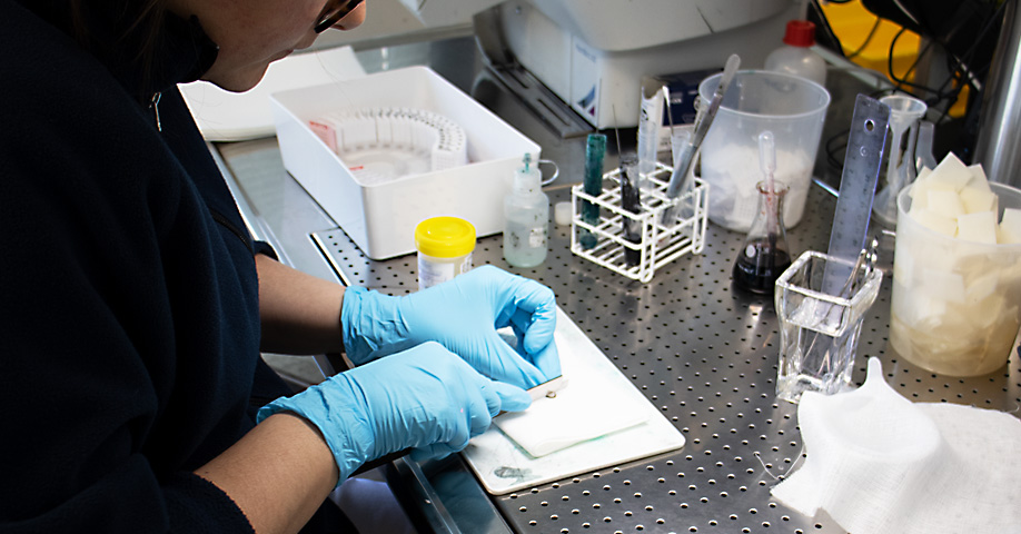
The role of our Biomedical Technician (BMA) is to take the samples, analyze them macroscopically before they are dissected to makes it easier to obtain surfaces that can be diagnosed by the pathologist in the last step. The role of the BMA is highly technical, requiring a great deal of knowledge and experience. 75% of the samples we receive are difficult skin samples that require expertise to dissect and handle. The sample ID label is scanned into the LIS and the case is automatically opened on the computer, the tissue is analyzed macroscopically and the BMA completes the case with a description of the preparation's appearance and the work steps performed by the BMA. The technician registers the number of pieces cut from the sample and automatically receives cassettes with printed sample and cassette ID on it. The sample pieces are placed in cassettes, logged in baskets and placed into the dehydration processor depending on the type of tissue, consistency and amount of fat.
Our laboratory has modern premises and modern equipment, which allows us to not handle large amounts of formalin on site, the formalin is poured into funnels. These lead down to a large tank (3 kbm) in the basement that collects all the formalin handled. This tank is emptied by Stena when our alarm signals that it has reached a certain level. This allows for the formalin levels within the laboratory to remain low for the safety of our laboratory technicians.
To be continued...
Steg två – Nedläggning
Alla vävnader läggs ned i formalin direkt när läkaren tagit ditt prov. Formalinet gör att alla levande processer stannar av så att alla celler i vävnaden ser exakt lika ut som om det vore kvar på kroppen. Om inte detta steg görs, ruttnar vävnaden och vi kan då inte se hur cellerna ser ut ”live” och då inte ge en bra diagnos. Steg två i labb-processen är nedläggning, där vi häller av formalinet från vävnadsprovet.
Våra Biomedicinska analytiker (BMA) tar ut proverna och analyserar dessa makroskopiskt för att skära dem i bitar som gör det lätt att få fram ytor som kan diagnosticeras av patologen i sista steget. Detta kräver stor kunskap och erfarenhet, speciellt då vi har 75% svåra hudar som man måste veta exakt hur de skall skäras och hanteras. Provets id-etikett skannas in och fallet öppnas automatiskt på datorn, vävnaden analyseras makroskopiskt och BMA kompletterar fallet med en beskrivning av preparatets utseende och de arbetsmoment som BMA utför. BMA anger hur många bitar respektive prov delas upp i och då skrivs det med automatik ut rätt antal kassetter märkta med provets och kassettens id. Provets delar läggs i kassetter som loggas in i korgar och körs i adekvat dehydreringsprogram beroende på typ av vävnad, konsistens och fettmängd.
Vårt laboratorium har moderna lokaler och modern utrustning och för att inte hantera formalin i större mängd på plats, hälls formalinen i trattar. Dessa leder ned till en stor tank (3 kbm) i källaren som samlar all hanterad formalin. Denna tank töms av Stena när vårt larm signalerar att den nått en viss nivå.
Fortsättning följer…